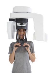
CBCT Scan In Bangalore
The introduction of Cone Beam Computed Tomography (CBCT) led to a change in the way Implantology and Oral and Maxillofacial Radiology is being practiced.
Radiation exposure dose from CBCT is 10 times less than from conventional CT scans. Furthermore, CBCT is highly accurate and can provide a three-dimensional volumetric data in axial, sagittal and coronal planes.
- Application in Dentistry
Replacement of missing teeth by dental implants demands accurate assessment of the implant site for the su
ccessful treatment outcome. It helps to avoid injury or encroachment of contiguous vital structures. Currently CBCT is the ideal choice, which has brought down implant failures by rendering accurate information about vital structures such as, height and width of the bone available, bone density and profile of the alveolus, while delivering low radiation exposure.
- CBCT provides pictorial guides for safe placement of mini-implants, evading accidental and irreparable injury to the vital structures. Surgical guide can be prepared with the help of the information retrieved using a CBCT which provides accurate guidance for placement of the proposed implants in various sites.
- Advantages of CBCT have made it the choice for exploring and handling midfacial and orbital fractures including dentoalveolar fractures, post fracture evaluation, interoperative visualization of the maxillofacial bones, and intraoperative navigation throughout procedures.
- Most important application of CBCT is to investigate the exact 3D location of jaw pathologies like benign or malignant tumors, inflammatory bone lesions, to assess impacted teeth, to investigate the exact location of supernumerary teeth and to assess their relation to vital structures.
- CBCT enables to examine the TM joint space and the true position of the condyle within the fossa, which is helpful in revealing likely dislocation of the joint disk.
- CBCT can be used to determine the number and morphology of roots and associated canals(both main and accessory), establish working lengths, and determine the type and degree of root angulation and as well provides true assessment of present root canal obturations.
- CBCT has been suggested for classifying the source of the lesion as endodontic or non-endodontic, which may influence treatment plan. Detecting vertical root fractures, measuring the depth of dentin fracture, and detecting horizontal root fractures comes handy due to absence of superimpositions and projection issues of 2D imaging.
In conclusion, CBCT is an advanced technology that is far better and safer than 2D radiographs for visualization of defects and fabricating treatment plans. It is the gold standard for implant planning.


